ABSTRACT
Intestinal enterococci are one of the two core microbiological parameters in the new drinking water legislation, used as indicators of faecal contamination. They must be detected in all analyses of drinking water. A total of 134 strains of enterococci (Enterococcus spp.) and 93 strains identified as background microflora, or as potentially false-positive strains, were examined from operational samples of treated (i.e. drinking) water. Untreated wells and boreholes, which are expected to carry a higher risk of faecal contamination, were not included. The most frequently detected Enterococcus species was E. casseliflavus (31 %), followed by E. faecium (25 %). The most frequent species not belonging to intestinal enterococci was Aerococcus viridans (n = 80); however, not all obtained strains survived the first passage. The lowest sensitivity to free chlorine was observed in E. hirae and also in the previously mentioned A. viridans. All strains were further tested using bile esculin azide medium (BEA test) after 2, 4 and 24 hours of incubation and for β-D-glucosidase (GLD) activity in a selective medium (Enterolert DW, IDEXX). A false-negative BEA test after 2 hours of incubation was recorded in 10 % of enterococci, most often in E. gallinarum, E. casseliflavus, and E. durans. Only 1 % of Enterococcus strains showed a false-negative results in the GLD test, but a further 7 strains (5.3 %) showed a weak reaction. A false-positive BEA test after 2 hours of incubation was recorded in 8 % of the background microflora strains, while a false-positive GLD test was observed in 14 % of the strains. The method according to the EN ISO 7899-2 standard is fully suitable for the detection of intestinal enterococci in drinking water. The use of alternative methods based on the determination of β-D-glucosidase activity is less appropriate, as it broadens the group of “intestinal enterococci” to include the entire Enterococcus genus, and the detection may therefore not clearly indicate faecal contamination.
INTRODUCTION
The detection of intestinal enterococci may appear straightforward at first glance, but it nevertheless has its pitfalls. On the one hand, intestinal enterococci are, in the new drinking water legislation, one of the two key microbiological indicators of faecal contamination, with the highest parametric value. On the other hand, alternative methods based on the detection of enterococci through β-D-glucosidase activity are gaining ground, extending the target group to all enterococci, i.e. Enterococcus spp. However, relatively little is known about the ecology of enterococci, particularly in drinking water, which complicates the interpretation of the results obtained. This study was therefore conducted, and the results were presented at the Vodárenská biologie 2025 conference [1]. A revised and extended version is presented here.
OVERVIEW OF THE ISSUE
Intestinal (faecal) enterococci are Gram-positive spherical or ovoid bacteria arranged in pairs or chains and belong to the genus Enterococcus (order Lactobacillales, phylum Firmicutes). Advances in molecular genetic methods in taxonomy have led to a continuous increase in the number of described enterococcal species; for example, only 19 species were known in 1995, whereas at present 60 species have been validly described [2]. With the now widely used MALDI-TOF method, individual species can be identified, allowing a more detailed interpretation of the results obtained. For example, among 101 isolates from various surface, technological, and drinking waters, the following species were identified: E. faecalis (26.7 %), E. hirae (20.8 %), E. faecium (18.8 %), E. casseliflavus (15.8 %), E. durans (11.8 %), and E. mundtii and E. moraviensis (both 2.3 %) [3].
Intestinal enterococci are regarded as indicators of faecal contamination, and their importance has increased in recent years – under the new legislation [4, 5], together with Escherichia coli, they represent a key indicator and must be determined in all types of analysis (both reduced and full). This is associated not only with an increased number of samples analysed, but also with a higher number of positive detections that need to be properly interpreted.
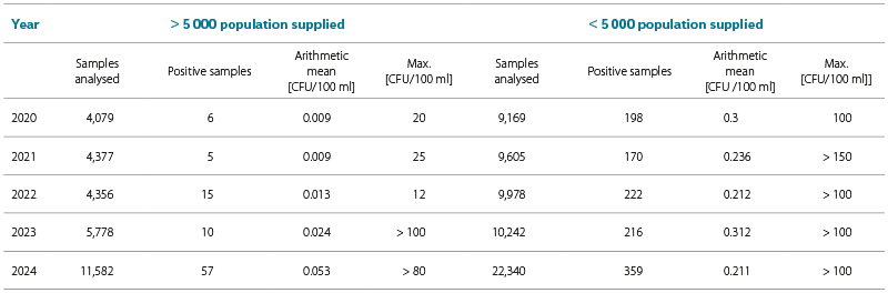
The frequency of detection in drinking water over the past five years in the Czech Republic, based on data from the National Institute of Public Health (Drinking Water Quality Report) [6], is presented in Tab. 1. At present, enterococci are the core parameter and its detection is included in all drinking water samples; consequently, the number of analyses, and thus the number of positive detections, is increasing (in Tab. 1, a marked difference can be seen particularly between 2023 and 2024). The higher numbers of detected enterococci are mainly associated with the increased number of tests performed. However, as the figures are still relatively low, it will be important to continue monitoring this situation.
Tab. 1. Detection of intestinal enterococci in drinking water in the Czech Republic (categories: > 5,000 population supplied, < 5,000 population supplied, total number of samples analysed, number of positive samples, arithmetic mean and maximum value) in 2020–2024 (CFU = colony forming units)
There is a substantial body of scientific literature on intestinal enterococci; however, it predominantly consists of studies describing the sources from which particular species have been isolated or the description and characterisation of new species. Where environmental studies do exist, they are primarily focused on surface waters and bathing waters, as well as on sludge and sediments. Studies dealing with a more in-depth investigation of enterococci isolated from drinking water are lacking. Thus, although numerous studies [7, 8] address the question of “who isolated which enterococcus, when, and from where”, relatively little is known about their actual ecology, particularly in relation to drinking water, drinking water treatment, and their survival and growth in biofilms.
Although all commonly identified enterococcal species occur in the intestines of humans or warm-blooded animals, a distinction is made between species that are typically faecal (E. faecium, E. faecalis, E. hirae, E. durans) and those associated with possible proliferation on plant material (E. mundtii, E. casseliflavus) [3]. Enterococci are also frequently used as indicators in microbial source tracking (MST), and various methods for their elimination from the aquatic environment have been described. They are also considered significant carriers of antibiotic resistance (e.g. to vancomycin and ampicillin). In the environment, they often occur in a non-virulent VBNC (Viable But Non Culturable) state.
For the determination of intestinal enterococci in drinking water, the long-established method according to ČSN EN ISO 7899-2 [9] is used. This method includes membrane filtration of samples, incubation for 48 hours at 36 °C on Slanetz and Bartley agar, and confirmation for 2 hours at 44 °C on bile esculin azide agar (hereafter BEA). Intestinal enterococci are defined as red to maroon colonies (Fig. 1) that, after subculture on the confirmation medium, show blackening of the medium beneath the colony. This method is intended to detect predominantly enterococcal species of faecal origin (E. faecalis, E. faecium, E. durans, and E. hirae). This is also related to the reduction of the confirmation time from four to two hours in 2001, based on the assumption that typical faecal enterococci exhibit faster and more intense BEA activity. More recent methods, which aim to serve as alternatives, are often based on the activity of the enzyme β-D-glucosidase, which enables the detection of all enterococcal species (Enterococcus spp.) [10]. This does not appear to be appropriate, particularly because this parameter is not merely an indicator but a key parameter with a limit value (unsurpassable parametric value). On the contrary, a more appropriate approach for improving the interpretation of enterococci results would be to move in the opposite direction, namely towards the identification of individual species.
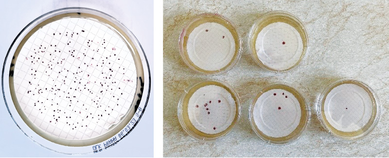
Fig. 1 a, b. Left: Aerococcus viridans, forming very small (mostly non-blackening) colonies; right: detection of intestinal enterococci
METHODOLOGY
This study included strains isolated from treated drinking water (but not from wells), obtained over a two-year period from operational hydroanalytical laboratories. The strains were purified, identified using the MALDI-TOF method (with the application of formic acid), and the confirmation test was repeated, with results recorded after 2, 4, and 24 hours. In addition, the strains were tested for β-D-glucosidase activity in a selective medium (Enterolert DW, IDEXX; more recently also according to ISO 7899-3 [11]).
Finally, representatives of the most frequently isolated species were tested for sensitivity to free chlorine using a method modified according to Annex 4 of Decree No. 409/2005 Coll. [12]. A solution of sodium hypochlorite was added to a measured volume (1,000 ml) of settled tap water at laboratory temperature to achieve a free chlorine concentration in the range of 0.15–0.17 mg/L. The solution was then artificially inoculated with the tested strains of the genus Enterococcus. The initial concentration of the test strain was approximately 10⁵ CFU/mL. Before the test, the contaminated water was thoroughly mixed (e.g. by shaking) to ensure uniform distribution of microorganisms. At test intervals of 1, 5, and 30 minutes, 0.5 ml of the prepared solution was inoculated onto the surface of a solid culture medium, and after incubation for 48 hours at (36 ± 2) °C, the colonies grown on the surface were counted. During the test period, the test solution in the flask was continuously mixed. Simultaneously, the original suspension was inoculated to determine the initial number of enterococci. All specified time intervals were tested; however, only the results after 1 minute were evaluated, as the results after 5 and 30 minutes were mostly negative. For each species, a strain isolated from treated drinking water and a strain isolated from the natural environment (bathing water) were tested in parallel, and all assays were performed in duplicate. After incubation, the relative reduction of the tested species (strain) after 1 minute of exposure to free chlorine was calculated in comparison with the control number.
RESULTS AND DISCUSSION
Identification of species
A total of 227 strains isolated in five water management laboratories from drinking (treated) water were processed; of these, 134 were subsequently identified as species belonging to the genus Enterococcus (a total of 10 species), while 93 belonged to other genera, with a clear predominance of Aerococcus viridans.
The species composition of enterococci and their relative distribution are shown in Fig. 2. The most frequently identified species was E. casseliflavus (31 %), followed by E. faecium (25 %), E. hirae (13 %), E. faecalis (10 %), and E. mundtii (9 %). Enterococcal species considered to be of faecal origin according to ČSN EN ISO 7899-2 [9] (faecalis, faecium, hirae, durans) accounted for only 54 %. For comparison, our earlier unpublished results from the identification of 612 enterococci from bathing waters showed that the most frequently identified species was E. faecium (25.2 %), followed by E. faecalis (21.1 %), E. durans (17.3 %), and E. casseliflavus (14.4 %). Such datasets can, of course, be compared only to a limited extent; nevertheless, it is evident that different matrices yield different results (unfortunately, the literature cited in the Introduction [3] analysed enterococci from a “mixture of matrices”). That bathing water represents a completely different matrix is also apparent from the composition of the accompanying microflora. According to our previous results, as well as the cited literature [3], the species most frequently interfering with the determination of intestinal enterococci in bathing waters was Lactobacillus plantarum, whereas in drinking water the most prevalent species of background microflora was Aerococcus viridans.
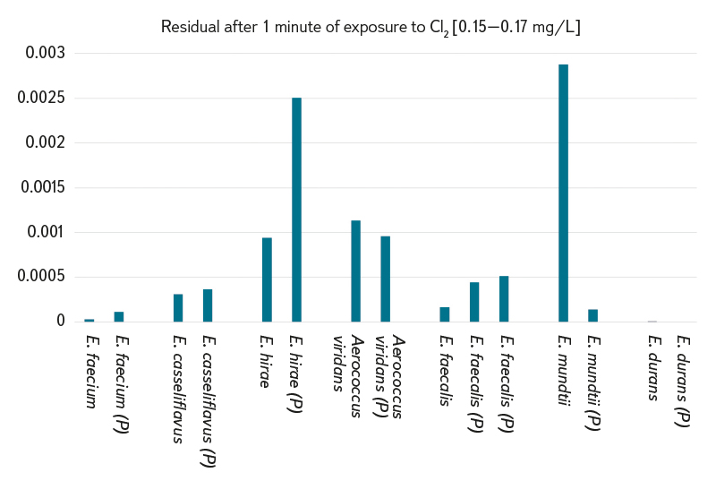
Fig. 2. Occurrence of individual species of intestinal enterococci isolated from drinking water
The high occurrence of E. casseliflavus in drinking water cannot yet be reliably interpreted; however, it should be noted that this was not a case of “many strains from a single sample”, as is often observed in bathing waters, but rather a more continuous occurrence. In previously cited studies, this species has often been associated with possible proliferation on plant material. In drinking water, the key question is how it behaves, for example, in biofilms or on sand filters, which is not yet known. In contrast to bathing water samples, where this species may proliferate, for example, on reeds and the membrane filter may then be covered with these (rather small) colonies, in this case overgrown filters were generally not observed. When membrane filters were covered with small colonies, these were exclusively A. viridans.
Confirmation and supplementary tests
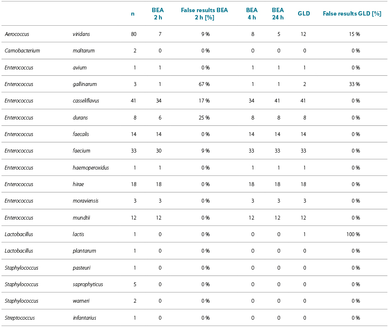
The results of the confirmation and supplementary tests for individual species are presented in Tab. 2. In addition to the absolute number of positive reactions for strains of individual species, the table also shows the relative proportion of false-positive results (in background microflora) and false-negative results (in enterococci).
According to the current version of ČSN EN ISO 7899-2 [9], the duration of the BEA test is 2 hours, which was taken as the reference time. Additional times (the previously used 4 hours and 24 hours) were tested for the purpose of further discussion of the results. However, the difference between 2 and 4 hours was minimal. A false-negative BEA test (i.e. after 2 hours of incubation) was recorded in 10 % of enterococci, most frequently in E. gallinarum, E. casseliflavus, and E. durans. A false-negative result in the β-D-glucosidase test was observed in only 1 % of enterococcal strains, but a further seven strains (5.3 %) showed a weak reaction. Unfortunately, it is not precisely known how a positive GLD test should appear, as no comparator is available for the Enterolert DW test. A false-positive BEA test (after 2 hours of incubation) was recorded in 8 % of strains of the accompanying microflora; in some strains of A. viridans, the positive reaction faded during the subsequent 20 hours. A false-positive β-D-glucosidase test was observed in 14 % of strains.
Sensitivity of enterococci to free chlorine
The most frequently isolated enterococcal species and the most common background species, A. viridans, were selected for testing their sensitivity to free chlorine. It is generally accepted that enterococci are less sensitive to the effects of free chlorine than, for example, coliform bacteria, and this is also confirmed by the results of our operational testing of disinfectants (commercial, unpublished data). For each species, two strains were tested (three strains in the case of E. faecalis), with one strain always isolated from treated drinking water (and
therefore potentially exposed to free chlorine) and the other from a typical natural environment (bathing water). According to legislative requirements [12], enterococci counts must be reduced by at least three orders of magnitude at a free chlorine concentration of 0.3 mg/L. The strains investigated in this study largely met this requirement even at half the concentration of free chlorine (0.15 mg/L). The results are illustrated in Fig. 3. The least sensitive species were E. hirae, A. viridans, and a strain of E. mundtii isolated from drinking water (rather than from bathing water), whereas the most sensitive species was E. durans. In 25 % of E. durans strains, a delayed BEA test reaction was observed (Tab. 2), which may be attributed to stress in strains originating from treated water. A certain degree of resistance to free chlorine in E. hirae may explain why this species ranked third among the identified strains, and a similar resistance in A. viridans may account for its frequent occurrence as accompanying microflora. The most resistant strain, E. hirae, is also prescribed for testing the bactericidal properties of disinfectants [13]. Until the final experiment, it appeared that strains from natural environments were more sensitive to the effects of free chlorine (possibly due to the absence of stress?) than strains isolated from drinking water; however, this was clearly not the case for E. mundtii, where the opposite was observed.
Tab. 2. Results of additional tests for strains of individual species; the number of strains examined (n), positive results of the BEA test after 2, 4 and 24 hours, positive results of the β-D-glucosidase (GLD) test. False results (false positives for background microflora or false negatives for enterococci) for BEA tests after 2 hours and GLD are given separately [%]
CONCLUSION
Although the “collection of enterococci” lasted at least two years, the number of strains obtained was not particularly high (enterococci = 134 + accompanying microflora = 93). Nevertheless, valuable data were obtained. The most frequently isolated species from drinking water was E. casseliflavus, which is usually less abundant and occurs rather sporadically in natural waters. This occurrence cannot yet be fully interpreted; one possible explanation is its ability to survive in biofilms (?). This species has also been associated with the potential for proliferation on plant material. Among the accompanying microflora, the most frequently identified species was A. viridans, which, together with E. hirae, showed the lowest sensitivity to free chlorine. The method according to ČSN EN ISO 7899-2 is fully suitable for the determination of intestinal enterococci in drinking water. The use of alternative methods based on the determination of β-D-glucosidase (GLD) activity is not entirely appropriate, as it expands the group of “intestinal enterococci” to include Enterococcus spp., thereby extending detection to species with an uncertain faecal origin. Moreover, the interpretation of enterococci results would benefit from the opposite approach, namely the identification of strains and interpretation of their occurrence in the environment. In addition, the most frequently detected species of accompanying microflora, A. viridans, shows false-positive GLD results in 15 % of cases. Given that this is a key parameter with the highest parametric value, such an expansion of the “group” could lead to complications.
Acknowledgements
This publication was supported by the Ministry of Health of the Czech Republic – Institutional Support (National Institute of Public Health – SZÚ, No. 75010330). Special thanks are extended to the staff of laboratories of water supply operators who provided strains for this study.
The Czech version of this article was peer-reviewed, the English version was translated from the Czech original by Environmental Translation Ltd.
